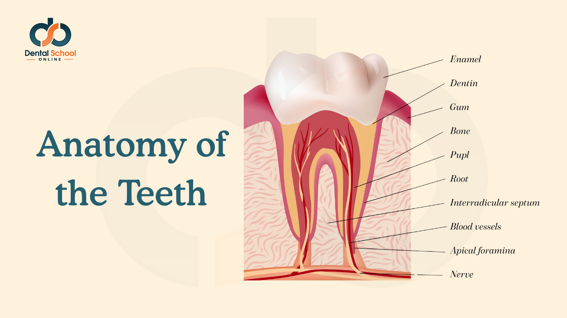
Understanding the anatomy of the teeth is essential for diagnosis, treatment planning, and clinical procedures. Each component has a specific structure and function, contributing to the overall health and function of the oral cavity.
📌 Key Components of a Tooth
Enamel: The hardest, outermost layer of the tooth, providing protection against wear and decay.
Dentin: A calcified tissue beneath the enamel, less hard than enamel, contains tubules that transmit sensations.
Pulp: Soft tissue in the center of the tooth containing blood vessels, nerves, and connective tissue.
Gum (Gingiva): Soft tissue surrounding the teeth, protecting roots and underlying bone.
Bone (Alveolar Bone): Supports and anchors the teeth in the jaw.
Root: Part of the tooth embedded in the bone, anchoring it in place.
Interradicular Septum: Bone located between the roots of multirooted teeth, supporting periodontal structures.
Blood Vessels: Supply nutrients and oxygen to the pulp and surrounding tissues.
Apical Foramina: Openings at the tip of the root through which nerves and blood vessels enter the pulp.
Nerve: Transmits sensation, including pain, from the tooth to the brain.
🦷 Tooth Layers
Layer / Structure
Description
Enamel
Hard, mineralized outer layer; protects dentin and pulp.
Dentin
Beneath enamel; sensitive tissue with tubules transmitting stimuli.
Pulp
Central soft tissue containing blood vessels, nerves, and connective tissue.
Root
Embedded in bone; anchors tooth and contains pulp canals.
Gum (Gingiva)
Protective soft tissue surrounding teeth and supporting bone.
Bone (Alveolar Bone)
Supports and anchors teeth in the jaw.
Interradicular Septum
Bone between roots of multirooted teeth, supports periodontal structures.
Blood Vessels
Supply nutrients and oxygen to pulp and surrounding tissues.
Apical Foramina
Openings at root tip allowing nerves and blood vessels to enter pulp.
Nerve
Transmits sensations including pain from the tooth to the brain.
🧠 Quick Learning Tips
Visualize layers from enamel → dentin → pulp → root for easier memory.
Use diagrams to connect anatomical terms with their locations.
Remember that blood vessels and nerves enter via apical foramina in the root tip.
📝 Mini Quiz
Test your knowledge! Answer in the comments below 👇
Which layer forms the hard, protective outer surface of the tooth?
Where are the blood vessels and nerves located inside the tooth?
What is the function of the interradicular septum?
⭐ Summary
The anatomy of the tooth is composed of multiple layers and structures, each with a specific role in protecting, supporting, and nourishing the tooth. Mastery of these components is crucial for clinical practice and understanding dental pathology.
Founder of Dental School Online, Dr. Ahmed combines years of dental practice and teaching experience to create high-quality, exam-focused online courses for dental students across the Gulf region.